

Open daily 9:00 AM — 8:00 PM, free parkingSevere pain — we try to see you the same dayPrice and warranty are fixed in a written treatment plan
Dentistry in Almaty
We treat under a microscope, plan in 3D, and document every stage with photo records. You always see what you're paying for.
30+
years treating patients in Almaty
up to 5
years of warranty on the implant — for other work, the term is specified in the contract
0%
instalments: Jusan bank — 24 months, Kaspi — 12
0 ₸
initial examination and treatment plan
Our approach
We save your natural teeth instead of selling implants
Replacing a tooth with a titanium implant is simpler and more profitable for a clinic. We take a different route: first we estimate how many years your own tooth still has, and only then discuss alternatives.
For complex cases we convene a case review, and sometimes invite colleagues from other clinics. That is normal practice when the goal is to treat rather than to fill a shift.
Services
Complete treatment cycle in one dental clinic
From the first diagnosis to the final restoration — no referrals to other centers.
Microscope-assisted therapyCavities, pulpitis, and root canal retreatment with up to 25× magnification. We preserve as much healthy tissue as possible.Implantology and surgeryStraumann, Biohorizons, laser protocol, All-on-4 and All-on-6 in a single visit.OrthodonticsDamon and American Orthodontics braces, aligners, bite correction for adults and children.Aesthetics and prostheticsE-max veneers, zirconia crowns, digital Digital Smile Design before treatment begins.Cavity treatmentLatest-generation composites, single-visit treatment, and pain-free care.Professional hygieneUltrasonic scaling, Air Flow, polishing, and remineralization in one visit.E-max veneersThin ceramics without grinding down healthy tissue where possible.Crowns and denturesZirconia and ceramic, removable and fixed restorations, implant-supported work.Braces and alignersMetal, sapphire, and lingual systems, clear aligners.Teeth whiteningIn-office ZOOM and take-home trays. We record the shade in the treatment protocol.Pediatric dentistryAppointments from age three, treatment in a playful format, fissure sealants.Gum treatmentPeriodontitis and gingivitis: pocket cleaning, laser therapy, maintenance care.
Our dental clinic's special offers


Microscope with 25× magnificationPhoto documentation of every stageCone beam computed tomography3Shape intraoral scannerWarranty up to 5 yearsClass B autoclavesSince 1995Microscope with 25× magnificationPhoto documentation of every stageCone beam computed tomography3Shape intraoral scannerWarranty up to 5 yearsClass B autoclavesSince 1995
About us
One of the oldest dental clinics in Almaty: since 1995 — and today with a microscope, a CT scanner and an intraoral scanner
We opened in 1995 and have grown from a two-chair practice into a full-cycle clinic with an in-house dental lab, CBCT imaging, intraoral scanning and a dedicated implant surgery suite.
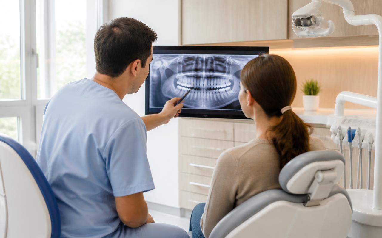
Six minutes on diagnostics, photo protocol and building a treatment plan
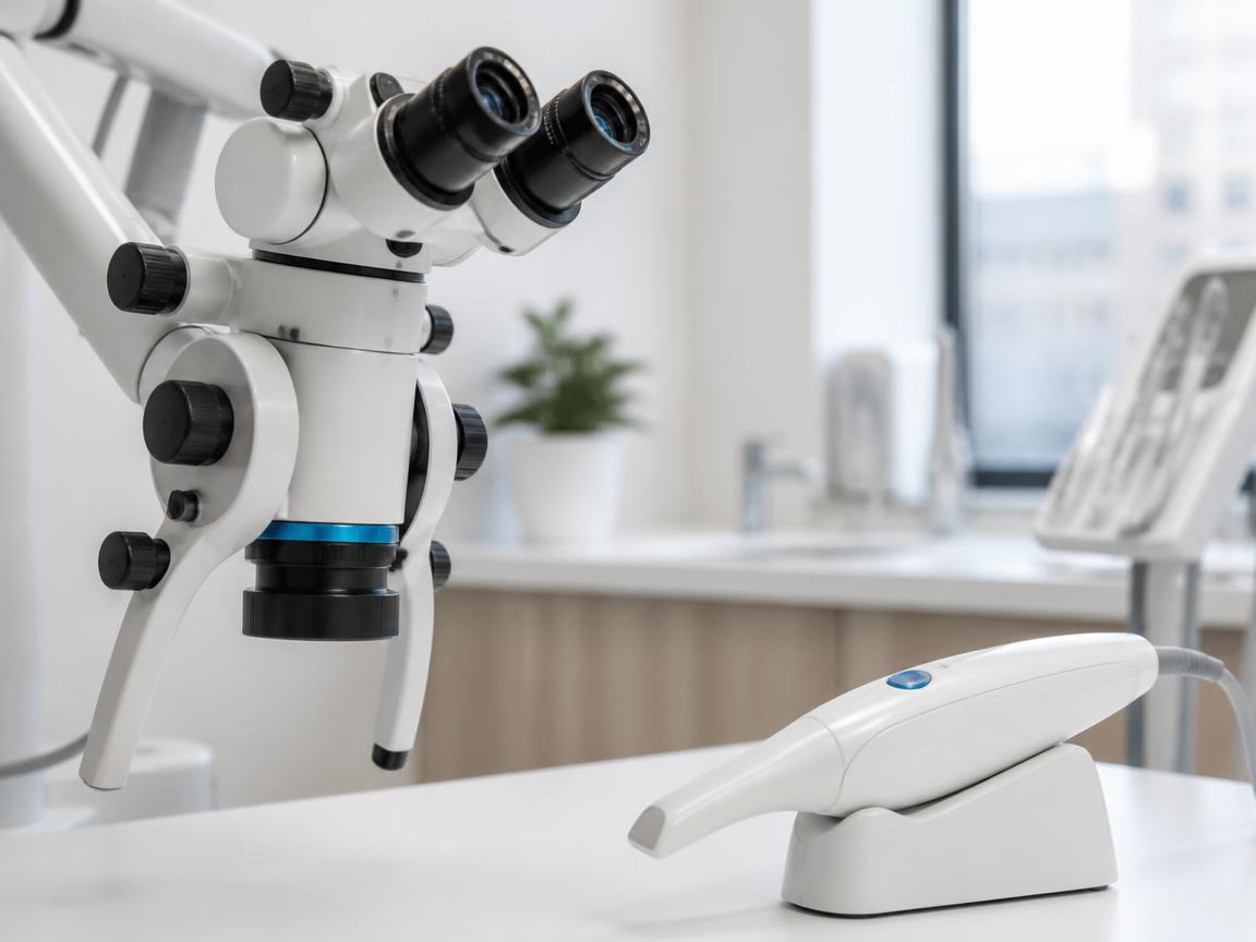
A 25× surgical microscope, cone-beam CT scanner, 3Shape intraoral scanner and a soft-tissue laser.
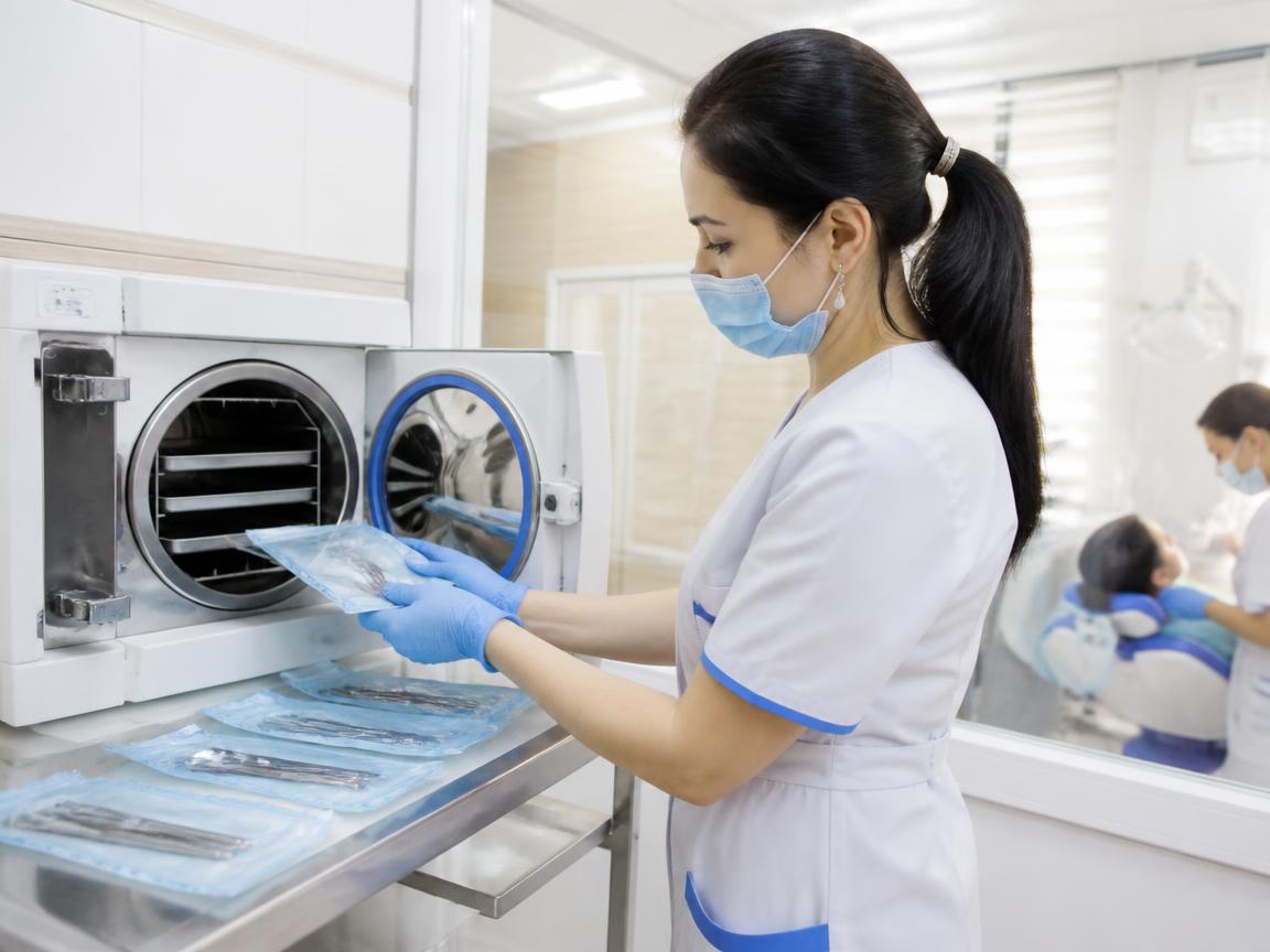
Class B autoclaves, single-use kits for every patient, a separate instrument processing cycle and indicator-based verification.
Principles
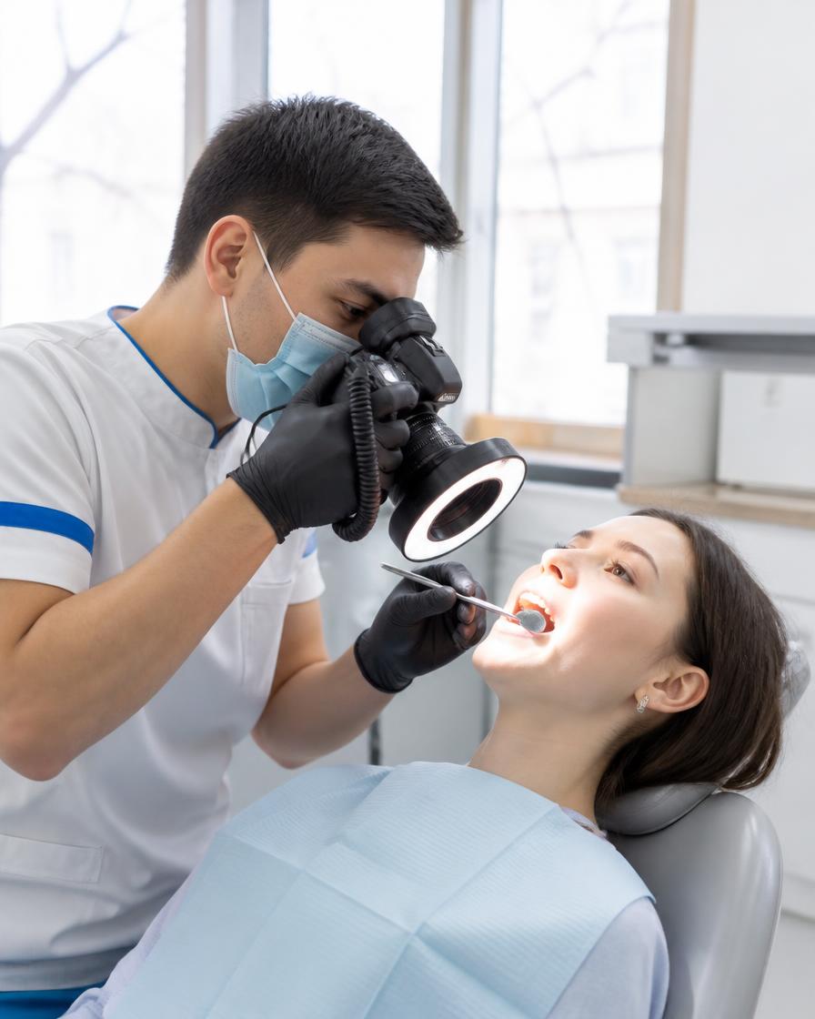
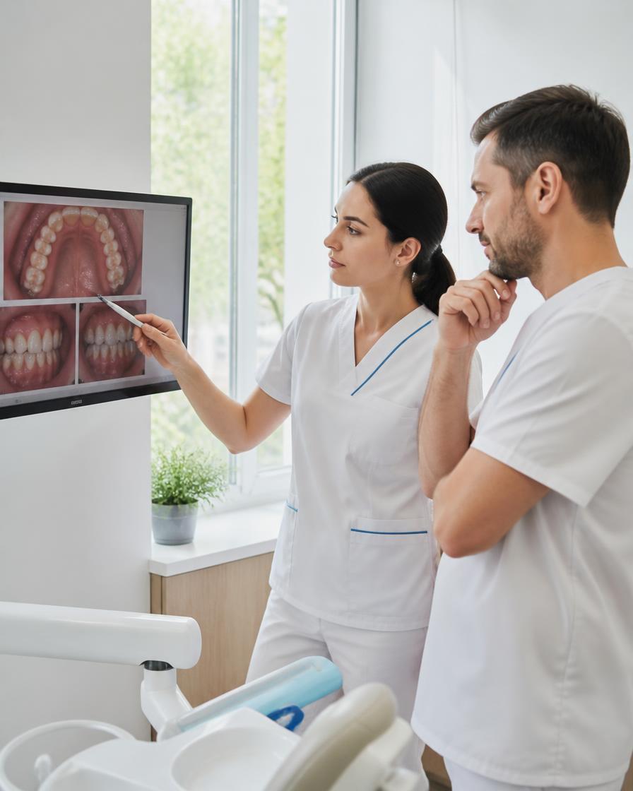
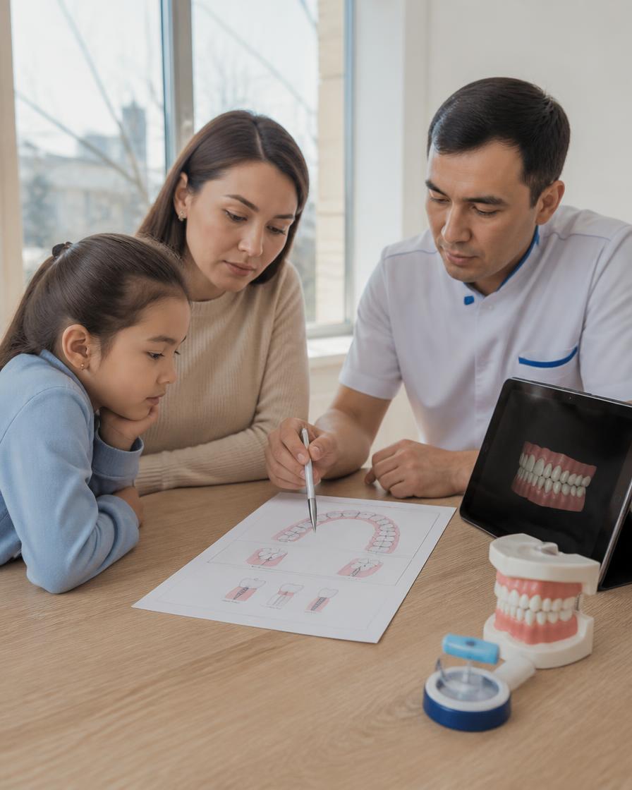
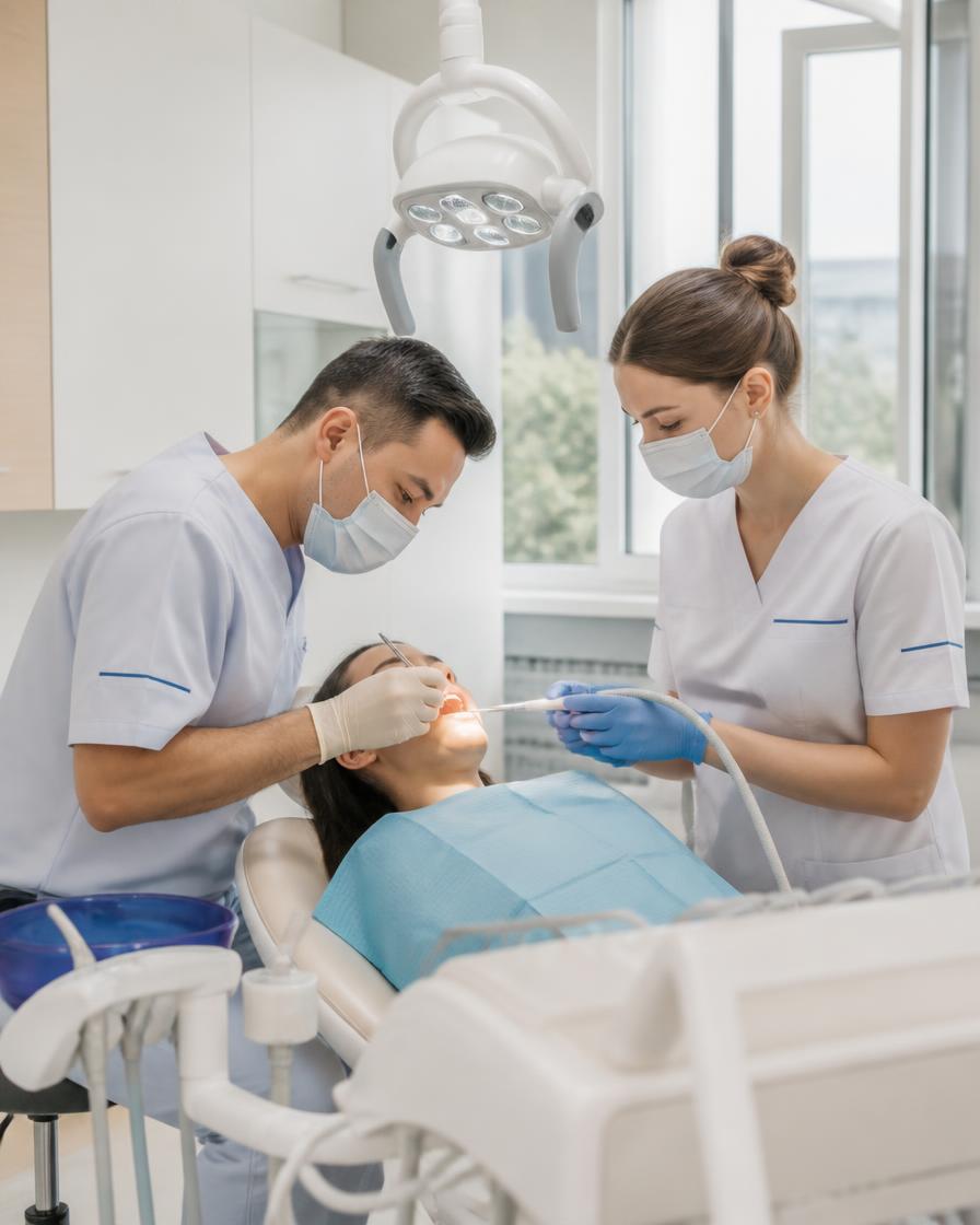
How treatment works at the Dental-Center dental clinic


Cost calculator
Get a preliminary cost estimate in 20 seconds
Which service do you need?
How many teeth are bothering you?
How would you prefer to pay?
One last step
Leave your contact details and we'll send the estimate and suggest a convenient time.
Request received
Our coordinator will contact you during clinic hours, daily 9:00 — 20:00.
Based on current 2026 pricingInstallment options with no extra chargesOur team replies during business hours: daily 9:00 AM — 8:00 PM
Prices
Full price listCost of treatment
The price is fixed in the treatment plan and does not change during the process.
TreatmentDuration & warrantyPrice
Prices are indicated in tenge and are valid from August 1, 2026.
Our work
Before and after
Drag the slider to compare before and after treatment.


Treatment of secondary caries and restoration of 4 teeth
We restored the natural fissures to preserve the bite. One visit, 2 hours. 5-year warranty.


Treatment of deep caries under a microscope
A small spot on the surface turned out to be an extensive cavity. We kept the tooth vital without root canal treatment.


Replacement of amalgam fillings with composite
Three posterior teeth in one visit: preparation, restoration of contacts, ridge contouring.


Treatment of pulpitis in three canals
Root canal filling under a microscope, restoration of anatomy, fissure sealing. 5-year warranty.


Treatment of 3 teeth in a single visit
Restoration of the posterior teeth: 4.5 hours of work, occlusal integration and final polishing.


Comprehensive professional hygiene
EMS ultrasound, Air Flow, brush polishing and application of a remineralizing gel.


Repair of chipped and darkened enamel
The enamel had an uneven shade with darkening, a chip, and plaque near the gum. Restoration and polishing were performed to restore uniformity.


Correction of tooth shade and shape
The enamel had an uneven shade with darkening, a chip, and plaque near the gum. Aesthetic restoration and cleaning were performed to even out the tone and contour.


Repair of chip and plaque
The enamel had darkening, a chip, and plaque near the gum. Aesthetic restoration was performed to restore a uniform shade.
How to choose a dental clinic?
Choosing a clinic isn't about the sign out front or a discount on implants. It's more about how the clinic handles diagnostics, sterilization, and communication with the patient.
What to look at first
- Diagnosis before treatment: an X-ray or CT scan, an examination, and a written plan. If the plan is only described verbally and you're immediately invited into the chair, that's a reason to ask questions.
- Sterilization: ask how instruments are processed. A class B autoclave covers the issue of cavities and narrow canals, and disposable supplies should be opened in front of you.
- Documents and license: private clinics operate under a license, and its details are usually displayed on a board. A contract, receipts, and a warranty card are the norm, not a favor.
- Team of doctors: each specialist has their own area — general dentistry, surgery, orthodontics, pediatric care. When one person performs all procedures, some of the work gets shortchanged.
- Reviews: read not the rating but the descriptions of situations. It's helpful when a person writes what exactly was done, how many visits it took, and how a disputed issue was resolved.
- Logistics: the commute to the clinic and its appointment schedule affect whether you'll see the plan through. A family clinic with daily hours is more convenient for a household of several people.
What to clarify at the consultation
- Treatment plan: which teeth are being treated now, which can be monitored, and what happens if treatment is delayed. Ask to see the problem on the X-ray — it makes it clearer what you are paying for.
- Who is managing the case: the name of the dentist responsible for the result, and who to contact if something bothers you after the appointment. This is especially important for implants and orthodontics.
- Stages and visits: how many appointments the plan involves and what determines that number. No one can give exact timelines in advance — the dentist decides based on the clinical picture.
- Warranty: what it covers, what is required to keep it valid (for example, hygiene visits and check-ups), and which cases are not covered.
- Payment: how the estimate is calculated, whether the amount can be split into payments, and what happens if the plan needs to be expanded during treatment.
- Emergencies: what to do at night or on a weekend if you have severe pain or a restoration comes loose. "Call us and we'll sort it out" is better than silence.
Why a single visit doesn't give the full picture
A single appointment shows the condition of the teeth and gums, but not how they will behave going forward. Some problems only become apparent over time: pocket depth, pulp response, implant healing, the rate of tooth movement with braces. That's why a clear plan is usually broken into stages, and after each one the doctor compares the actual result with what was expected. It also happens that the picture looks one way during the exam and is refined after a CT scan or under the microscope — that's normal diagnostics, not a mistake. Hence a simple rule: don't rush to sign up for a large amount of work on the very first day if no one has explained to you why it's done that way and what will happen at each step. And don't choose a clinic based on a single good review — look at how follow-up visits, reminders, and complaint handling are organized. It's boring, but it honestly shows who you're dealing with.
How pulpitis and periodontitis differ and how they are treated
Both diagnoses involve inflammation, but the focus lies at different depths. This determines the symptoms, the imaging, and the extent of the intervention. Below we'll look at where the boundary between the conditions lies and how treatment is structured.
Where the boundary of inflammation lies
The pulp is loose tissue with blood vessels and nerves inside the crown and root canals. As long as the process is confined to it, it's called pulpitis. The tooth aches and reacts to cold and hot, and the pain radiates to the temple or ear. Sometimes it subsides on its own, and the person puts off the visit. That's a mistake. Next, the inflammation spreads beyond the root apex into the periodontal ligament — the tissue that holds the tooth in the bone. That's how periodontitis begins. Here the pain is different: dull, aching, worse when biting down, and the tooth feels as if it has grown and become taller than the others. The gum above it swells, and a fistula may appear. On an X-ray, a shadow is visible at the apex — an area of bone loss. The boundary between the diagnoses runs exactly at the root apex. Both conditions are treated by a general dentist, but with periodontitis the amount of work and the number of visits are greater. At the appointment, the doctor first takes the history, then takes an X-ray and performs tests. Only after that is an accurate diagnosis made.
Comparison of the two diagnoses
| Feature | Pulpitis | Periodontitis |
|---|---|---|
| Location of the source | Inside the tooth, in the pulp | Beyond the root apex |
| Nature of pain | Paroxysmal, triggered by temperature | Dull, on biting |
| Reaction to cold | Sharp, prolonged | Usually mild |
| X-ray | Often no changes | Shadow at the root apex |
| Gum | Calm | May swell, fistula |
| Scope of treatment | Root canals, filling | Root canals, retreatment |
| Number of visits | One to two | More often two or more |
How treatment is structured
- Diagnosis: the dentist examines the tooth, tests its reaction to cold and biting, and takes an X-ray. If the picture is unclear, a cone-beam CT scan is ordered to see the lesion in three dimensions.
- Anesthesia: local anesthesia is administered, and all subsequent steps are painless. The patient feels only touch and pressure, not sharp twinges.
- Access to the canals: the doctor removes the affected tissue, opens the tooth chamber, and locates the canal orifices. A microscope with 25× magnification helps, revealing what the naked eye cannot see.
- Canal treatment: the canals are shaped with instruments, irrigated with solutions, and filled down to the apex. In cases of periodontitis, medication is sometimes left in place for several days and a follow-up visit is scheduled.
- Filling: after the canals are sealed, a temporary filling is placed, followed by a permanent one. If the tooth walls are severely damaged, a crown is discussed — but that is a separate matter.
- Follow-up: a few months later, an X-ray is taken to check how the tissue is healing. If the lesion has shrunk, the tooth is kept under observation; if not, re-treatment is considered.
- What affects the outcome: the depth and duration of the process, the condition of the root, and the quality of canal treatment. An accurate prognosis is given by the doctor after examination and X-rays; it is not stated in advance.
Why professional oral hygiene is needed
A toothbrush at home removes plaque from smooth surfaces. Calculus, pigment, and deposits under the gum remain. Professional cleaning removes what home care can't.
What the dentist removes and what's left for home
A person removes soft plaque themselves if they brush regularly and correctly. After a day it mineralizes and turns into calculus. Calculus sits firmly and can't be removed with a brush. The dentist uses an ultrasonic scaler: it breaks up the deposits and washes them away with water. Pigment from coffee, tea, and tobacco is removed with air abrasion, and then the surface is polished with paste. Polishing matters: smooth enamel collects new plaque more slowly. Biofilm accumulates under the gum, causing inflammation and bleeding. It is removed with curettes or a laser. What's left for home is daily work: brush, toothpaste, floss, interdental brushes, water flosser. None of these remove calculus, because calculus is a hard structure, not a film. Prevention doesn't replace treatment, but it reduces the risk of gum inflammation. The dentist decides: some patients need a cleaning once a year, others need more frequent monitoring.
Stages of the procedure
- Examination: the dentist checks the gums, tooth mobility, the condition of the enamel and old fillings, and orders an X-ray if needed.
- Anesthesia: if sensitivity is high or deposits are deep under the gum, a gel is applied or an injection is given so the procedure goes smoothly.
- Ultrasound: the tip breaks up tartar above and below the gum line, water rinses away the broken-off particles, and plaque comes off the enamel without pressure.
- Air Flow: a mixture of air, water, and powder removes soft plaque and pigment in hard-to-reach areas, including the spaces between teeth.
- Polishing: a paste smooths the enamel so that plaque does not settle quickly and the surface stays smooth to the touch.
- Fluoridation: a coating strengthens the enamel and reduces sensitivity after the procedure, especially if heavy tartar was removed.
- Recommendations: the dentist shows which products to use for home care and schedules the next visit based on the condition of the gums.
Who needs it and how often
The frequency depends on the clinical picture. A person with healthy gums and good hygiene only needs to come once a year. If there is bleeding, periodontal pockets, crowded teeth, or braces, the interval is shortened. Smokers and coffee lovers need cleaning more often: pigment and plaque accumulate faster. With diabetes and certain medications, gums react more strongly, so monitoring is stricter. Pregnant women should have cleaning in the second trimester: hormonal changes alter gum response to plaque. For children, cleaning is done as indicated, usually after permanent teeth appear. The exact interval is determined by the doctor after examination. If gums bleed during cleaning, this is not a reason to skip hygiene — on the contrary, it's a signal to schedule an appointment. Professional cleaning does not treat cavities and does not replace treatment of pulpitis. It removes the cause of gum inflammation and helps the doctor see problems at an early stage. It depends on the case: sometimes after cleaning, additional gum therapy is needed, and the doctor decides this.
How aesthetic tooth restoration is done
This is the restoration of tooth shape and color with composite directly in the mouth. The doctor applies the material layer by layer and sculpts it by hand. The method requires a dry field and precise work.
When composite is appropriate
Composite restoration is chosen for enamel chips, worn edges, cracks without nerve damage, gaps between teeth, and darkened fillings in the visible area. The material is built up if the neighboring teeth are healthy and do not require crowns. The indication is determined by the doctor based on X-ray and examination: sometimes inflammation is hidden under a seemingly small defect, and then it is treated first. Such work does not replace orthodontics if the cause of chips is malocclusion. In these cases, the dental arch is aligned first, otherwise the composite chips again. The limitation is large destruction: when the tooth walls are lost, a crown is stronger. A separate topic is color: selecting the shade is harder than it seems, especially on front teeth where light falls at different angles. The patient should understand that even careful work darkens at the edge over time, and this is normal, not a defect.
Step-by-step process
- Examination and imaging: the dentist assesses the depth of the defect, the condition of the nerve and adjacent teeth, and if necessary takes an image with a cone-beam CT scanner.
- Shade selection: the shade is chosen in daylight by matching the enamel of the adjacent teeth, sometimes with a photo protocol, to achieve a seamless blend of shades.
- Isolation: the working field is dried with a rubber dam; saliva and moisture from breathing interfere with the bonding of the composite to the tooth tissue.
- Preparation: the affected tissue is removed, and a bevel is shaped on the enamel along the edge — it is needed for a smooth transition of the material into the tooth.
- Etching and adhesion: the enamel and dentin are treated with acid and bonding agent, which chemically bonds the composite to the tooth.
- Layered application: the material is applied in thin layers, each cured with a lamp; this is how the shape, translucency and color transition are created.
- Modeling and finishing: the cusps and edges are restored, then the restoration is ground and polished — this determines whether the work will be noticeable.
What the doctor decides
The outcome depends on the clinical picture. The doctor decides whether composite is enough or a crown is needed, whether depulpation can be avoided, and which shade to use as a base. Front teeth are more difficult: not only shape matters, but also how the material transmits light. Sometimes the defect affects the gum, and then inflammation is first removed, and restoration is postponed. The prognosis for service life varies: for some, the work lasts for years, for others, a chip appears earlier, and this is related to bite, habits, and hygiene, not just the material. The patient can influence the result: do not bite nails or pens, do not open packages with teeth, and come for check-ups. If the composite darkens or chips at the edge, it is updated or redone — this is repairable work. The doctor separately explains that polishing is not eternal: over time, the surface dulls, and it is refreshed. The decision on tactics is always up to the specialist, not the price list or the desire to do it quickly.
What All-on-4 is and when it is indicated
The All-on-4 protocol is a method of full restoration of the dental arch on four implants. Let's break down what the essence is, who it suits, and how preparation goes.
The essence of the protocol
Four implants are screwed into the jaw: two vertically in the anterior region, two at an angle in the posterior regions. The angulation provides support where there is little bone and, in some cases, makes it possible to avoid bone grafting. A bar or a custom framework is placed on the implants, and a fixed prosthesis is placed on that. It rests on the implants rather than on the gum, so it does not shift during chewing and does not press on the mucosa. The chewing load is distributed differently than with a removable denture. The position of the supports is planned based on cone-beam computed tomography and intraoral scanning data. If a preliminary dental consultation is needed, it is performed before the tomography in order to assess the general condition of the oral cavity. The protocol is used more often on the lower jaw, and on the upper jaw — according to indications. The outcome depends on the clinical picture: bone volume, gum condition, and concomitant diseases. The doctor decides after the examination and imaging.
Indications and limitations
| Situation | All-on-4 | Comment |
|---|---|---|
| Complete edentulism | Indicated | Restores the entire arch |
| Terminal defects | Possible | Determined by the doctor based on imaging |
| Severe bone atrophy | Limitation | Bone grafting is sometimes required |
| Diabetes mellitus | Limitation | Requires glycemic control |
| Smoking | Limitation | Increases the risk of complications |
| Bruxism | Limitation | A protective night guard is needed |
How preparation is carried out
- Examination and diagnostics: the doctor evaluates the oral cavity, performs cone-beam computed tomography and an intraoral scan to assess bone volume and nerve position.
- Surgical planning: based on the imaging, the implant placement sites and angulation are selected, the future prosthesis is modeled, and blood tests (glucose, coagulation) are ordered if necessary.
- Oral sanitation: caries, pulpitis, and gum disease are treated, and teeth that cannot be preserved are extracted to reduce the risk of infection after surgery.
- Preoperative preparation: a few days before the procedure, medications affecting coagulation are discontinued, the patient is prescribed antibiotics and rinses, and dietary recommendations are given.
- Implant placement: the surgery is performed under local anesthesia or sedation, takes several hours, and a temporary prosthesis is fixed the same day or a few days later.
Which is better: braces or aligners?
There is no clear winner. The choice depends on the clinical picture, age, and willingness to follow the rules. Below is how the systems differ and what the doctor weighs.
Differences in design and discipline
Braces are a fixed appliance: brackets are bonded to the teeth and connected by an archwire that gradually guides the teeth into position. You cannot remove it yourself. Aligners are removable transparent trays that are changed according to a schedule, usually every one to two weeks. This is the main practical difference. Braces work continuously, while trays work only while they are worn. If they are worn less than prescribed, tooth movement slows or stops, and treatment then takes longer. That is why with aligners, discipline matters more: they are removed for eating and brushing, and the rest of the time they stay in the mouth. There are also everyday differences. Trays are almost invisible and do not injure the mucosa, but they are easy to lose or forget to put on. Braces are visible and require more thorough hygiene around the brackets, but they do not depend on the patient's memory. Which is better in a particular case is decided by the doctor after the examination and imaging.
Comparison by characteristics
| Feature | Braces | Aligners |
|---|---|---|
| Removability | Fixed | Removable |
| Visibility | Visible | Almost invisible |
| Discipline | Does not depend on the patient | Depends on wear time |
| Hygiene | More difficult around brackets | Easier, the aligner is removed |
| Complex cases | Broader capabilities | Depends on the clinical picture |
| Loss of the appliance | Not possible | The aligner can be lost |
What the orthodontist takes into account
- Case complexity: in severe skeletal discrepancies and tooth rotations, the capabilities of the systems differ, and the choice is dictated by the clinical picture rather than the patient's preference.
- Age: in children and adolescents, tooth replacement and jaw growth introduce adjustments; in adults, the condition of the periodontium and roots is taken into account.
- Willingness to wear aligners: if the patient removes them more often than prescribed, the result is questionable, and the doctor may suggest a fixed appliance.
- Hygiene and enamel condition: plaque and gum inflammation are resolved before starting, otherwise treatment is paused; preparation before orthodontics is carried out in advance.
- Diagnostics: the plan is built on imaging and scans, so a cone-beam computed tomography scanner and a 3Shape intraoral scanner are required.
- Visit schedule: follow-up appointments are scheduled on weekdays, but if necessary, an appointment is possible on Sunday if the clinic operates without days off.
How do zirconia crowns differ from veneers?
Both options cover the tooth on top, but they solve different problems. Let's break down where the line is and why sometimes the choice is obvious, and sometimes the doctor decides.
Different tasks and different preparation
A crown covers the tooth entirely, on all sides, including the chewing surface and the cervical area. A veneer is a thin plate that is bonded only to the front, visible surface. Hence the different preparation: for a crown, more tissue is removed all around; for a veneer, only a layer from the front side, sometimes within the enamel. If a tooth is severely damaged, a single onlay cannot restore it: support is needed to hold the shape and protect against fracture. If the tooth crown is intact but the color or shape is unsatisfactory, a veneer makes more sense — it preserves more of your own tooth. A devitalized tooth, a tooth after root canal treatment, a tooth with a large filling — that's crown territory. Enamel chipping, a diastema, discoloration — that's veneer territory. The boundary is determined not by desire but by the condition of the tissues. A crown can also be placed on a molar; a veneer is not placed there: it won't withstand the load. Cost is not a selection criterion in this case, because redoing it costs more.
Key differences
- Coverage area: a crown covers the entire tooth, while a veneer covers only the front surface, so the amount of tooth reduction differs between them.
- Indications: a crown is placed on damaged and root-canal-treated teeth, while a veneer is placed on intact teeth when changing color or shape.
- Material: zirconia is opaque and strong, while veneer ceramic is thinner and translucent, which accounts for the difference in how color is rendered.
- Load: a crown withstands chewing pressure, while a veneer is not used on back teeth — it will chip or come off over time.
- Reversibility: tooth reduction for a crown is irreversible, while for a veneer the enamel is sometimes preserved, but this too is determined by the clinical picture.
- Removal: a crown is removed when redoing the work, and so is a veneer, but re-cementation is not always possible — it depends on the condition of the tooth.
What suits front teeth
At the front, not only strength matters but also how the tooth transmits light. Zirconia is denser, so a single crown made of it may look duller than neighboring teeth, especially near the gum. A veneer is thinner and works with translucency more naturally when it comes to color and shape. But a veneer has its limits: severe damage, a crack, root canal treatment — and it's no longer support. Then a crown is placed, sometimes with a ceramic facing over the framework to restore the liveliness of color. What will suit a specific case is decided by the doctor after examination and imaging. Sometimes the solution is mixed: some teeth are covered with veneers, others with crowns. Being close to home is convenient logistically, but the choice of restoration does not depend on the address — it depends on the clinical picture.
What equipment is used in the clinic?
Equipment determines how accurate the diagnosis will be and how precise the work is. Let's look at which devices are used at different stages — from imaging to instrument sterilization.
Diagnostics: from X-ray to CT scan
Diagnostics begins with an examination, but technology often makes the difference. A periapical X-ray shows a single tooth or a small area. Cone beam CT provides a three-dimensional image of the jaw: it is used to assess bone thickness, the position of canals and roots, and to plan implant placement. An intraoral scanner takes a digital impression — instead of a tray with paste, the dentist moves a small camera over the teeth, and a 3D model appears on the screen. It is needed for crowns, veneers, aligners, and orthodontic appliances. A microscope with 25× magnification helps to see what the naked eye cannot: canal orifices, enamel cracks, the margins of a filling. Some methods cannot be performed without special equipment, and the patient is referred elsewhere. Digital data is stored in the system and is available to the dentist at any stage — this is convenient when treatment is spread out over time. The dentist decides: not every case requires a full set of tests, sometimes an X-ray and an examination are enough.
Technology in treatment and prosthetics
- Microscope with 25× magnification: used in endodontics and root canal retreatment — magnification helps work more precisely in hard-to-reach areas, but whether it is needed depends on the specific tooth.
- Laser: used for soft tissue treatment, for example minor gum contouring; the method is not universal, and the decision remains with the doctor.
- Intraoral scanner: takes a digital impression for crowns, veneers, and aligners, replacing impression material and reducing the number of try-ins.
- Cone beam CT: provides a three-dimensional picture before implantation and complex prosthetic work, when planning from a regular X-ray is risky.
- CAD/CAM milling: crowns and inlays are milled from a digital model — this is a standard fabrication method, not a feature of an individual clinic.
- Articulator: reproduces the movements of the lower jaw to check the bite of the future restoration before it is placed and to avoid redoing it at the appointment.
Sterilization and anesthesia
Sterilization is the part of the work the patient does not see, but it is precisely what determines safety. Instruments go through several stages: disinfection, cleaning in an ultrasonic bath, packaging, and processing in a class B autoclave. Such an autoclave creates pressure and temperature that kill microorganisms, including spore forms. The packaging is opened in front of the patient — this is a simple sign that the cycle has actually been completed. Anesthesia is selected based on health status and the duration of the procedure. For topical anesthesia, a gel or spray is enough; for treatment, injectable drugs are used. The patient can ask for a pause at any moment: if sensitivity returns, the dentist will add more anesthetic. If you are looking for affordable dentistry in Almaty, do not sacrifice sterilization for the sake of price — instrument processing is not something to cut costs on. A separate topic is allergy to anesthetics: it must be mentioned before treatment begins, so that the drug can be chosen differently. It depends on the clinical picture and medical history.
What to keep in mind
Diagnostics matter more than advertising
An X-ray and an examination give more than promises on a banner. Cone beam CT shows bone volume, the position of canals, hidden lesions — things that cannot be seen with the naked eye. A microscope with 25× magnification helps to spot a crack or a remnant of filling material in a canal. An intraoral scanner takes a digital impression without a tray and plaster: fewer remakes at the fitting stage. Without this data, any treatment is built on guesswork. The patient should ask what the proposed plan is based on: which images were taken, what they show, and why this particular option was chosen. If the dentist answers substantively and shows the images on the screen, that is a good sign. If the conversation is only about discounts and promotions, that is a reason to be wary. Diagnostics takes time, sometimes more than one visit, but it is precisely what determines what happens next.
The treatment plan is discussed before it begins
The order of stages, their sequence, and the alternatives are discussed in advance. What is treated first: inflammation, then the prosthesis, or the other way around. Where a pause is possible and where delaying is not an option. What options exist if the budget is limited, and how one path differs from another in terms of the number of visits and the scope of intervention. A written plan is convenient: you can return to it, check it, and ask questions. A verbal agreement is forgotten, and details are later remembered differently. It is worth clarifying what is included in a stage and what counts as separate work. Separately — what the result will look like: shape, color, bite. If something changes along the way, that is also discussed and documented, not decided silently. The patient has the right to take a pause to think: haste in planning rarely does any good.
Prevention reduces the scope of intervention
Brushing twice a day, interdental brushes or floss where the toothbrush can't reach, and a check-up every six months. This isn't a guarantee that problems won't happen at all. But advanced plaque and small cavities are easier to treat than pulpitis or periodontitis. Early stages often avoid drilling: remineralization, fissure sealing in children, monitoring. Once the process reaches the nerve, we're talking about a different scope of work. Smoking, sugar, nighttime teeth grinding are factors that speed up destruction, and you should tell your doctor about them. Professional hygiene removes tartar and plaque, but doesn't replace home care. Visit intervals and the extent of intervention depend on the clinical picture: one person may just need an exam, another may need a plan for months. The doctor decides after diagnostics.
FAQ
Frequently asked questions
Initial examination, consultation with a specialist, and a written treatment plan — 0 ₸. If a CT scan or X-ray is needed, the administrator will tell you the cost of diagnostics in advance.
In most cases, yes. For cavities, chips, or acute pain, the doctor performs an examination, diagnostics, and starts treatment during the same visit. Complex surgical and prosthetic work requires preparation.
Call or message us and let us know the pain is acute. We keep reserve slots for urgent cases and try to see you on the day you contact us.
For therapeutic work and restorations — up to 5 years; for prosthetic structures and implants — according to the manufacturer's terms and protocol. The warranty is documented in writing along with the treatment plan.
Yes. Jusan bank — 24 months, Kaspi — 12 months. It takes about 10 minutes to set up right at the clinic, and no down payment is required.
Yes, a separate specialist handles pediatric appointments. Examinations, cavity treatment, prevention, and orthodontics — tailored to age and as gentle as possible.
Yes, for both adults and children. A consultation with an anesthesiologist and a medical examination are required before the procedure — these are arranged by the doctor at the initial appointment.
Our doctors and front desk staff speak Kazakh and Russian.
Reviews
All reviewsWhat our patients say
5,0
9 reviews on the site
9 ratings
ААйгерим К.12 August 2026
★ 5,0I've been afraid of dentists since childhood. Here the doctor first showed everything on the screen, explained every step, and only then started treatment. Two hours flew by without me noticing.
Cavity treatment
ДДанияр С.3 August 2026
★ 5,0I had four implants placed in a single visit. A week later I'd already forgotten I'd had surgery. Special thanks for honestly talking me out of unnecessary work.
All-on-4 implants
ММария В.28 July 2026
★ 5,0I had E-max veneers done. First they showed me a digital mock-up of my smile, I made some adjustments, and only then did they start the work. The result is exactly like the mock-up.
E-max veneers
ЕЕржан Т.19 July 2026
★ 4,9I bring my whole family here, including my 6-year-old child. The pediatric dentist found an approach within five minutes, and the treatment went without tears.
Pediatric dentistry
ААсель Н.5 July 2026
★ 5,0The braces were removed a month ahead of schedule. At every visit, progress was photographed, and you could see how everything was changing.
Damon braces
ННурлан Ж.22 June 2026
★ 5,0Came in with a dental abscess on a Saturday evening — was seen within forty minutes. They opened it, placed a drain, and explained what would happen next. By Monday the swelling was gone.
Emergency care
ГГульмира А.14 June 2026
★ 4,8For a year I couldn't get used to the removable denture from another clinic. Here they took new impressions with a digital scanner and remade it — the difference is huge.
Prosthetics
ССанжар Б.2 June 2026
★ 5,0I liked that the treatment plan was given to me printed out, with the price for each tooth. The final total matched the plan to the last tenge.
Comprehensive treatment
ТТогжан М.21 May 2026
★ 5,0I had a cleaning done. They showed me on the camera exactly where I wasn't cleaning well and gave specific recommendations, not just generic advice about flossing.
Professional cleaning
Leave a review
Tell us how your treatment went. The review appears on the site once the front desk has checked it.

Contacts
Always here for you
Not sure whether you need an appointment? Message us — the doctor will look at a photo and tell you whether it's urgent or can wait.
Message on WhatsAppWe reply during clinic hours, 9:00 — 20:00TelegramSend us a photo of the problem+7 747 093 89 86Daily, 9:00 AM — 8:00 PM, Open every day
AddressSatpayev 133/6, JAZZ Residential ComplexOpening hoursDaily, 9:00 AM — 8:00 PMParkingFree for patients
Blog
Useful materials


Contacts
How to find us
AddressAlmaty, JAZZ Quarter Residential Complex,
133/6 Kanysh Satpayev St.
133/6 Kanysh Satpayev St.
Phone+7 747 093 89 86
Opening hoursDaily, 9:00 AM — 8:00 PM
Open every day
Open every day
ParkingFree for patients
in the courtyard of the residential complex
in the courtyard of the residential complex
133/6 Kanysh Satpayev St.